Assessment and measurement
3D gait analysis
Cameras and force plates record how a person actually walks, joint by joint. It turns 'the walking looks better' into a measured before-and-after. Its clinical value is finding the cause of a limp — hip weakness, ankle stiffness, pain or leg length — and separating a compensation from the primary problem.
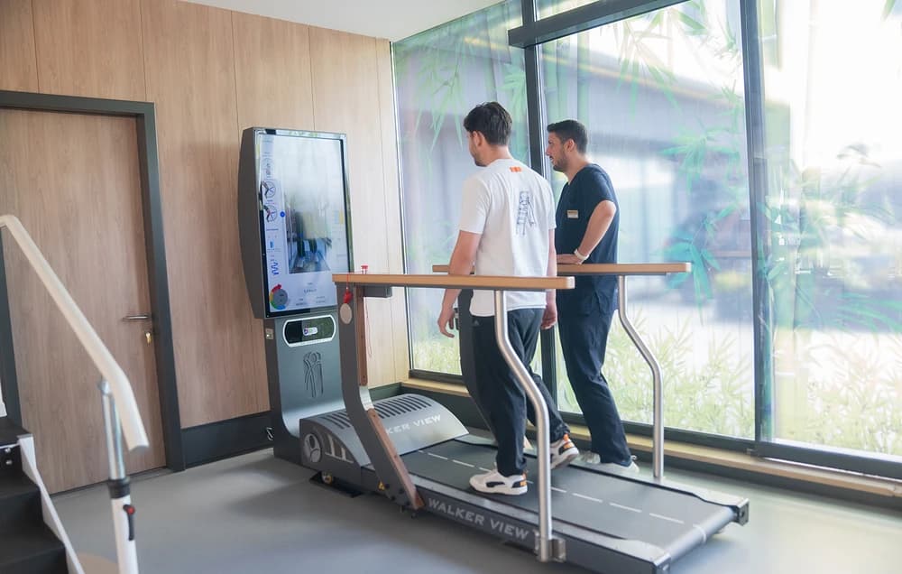
What instrumented gait analysis actually produces
Cameras track reflective markers on the body while force plates measure ground reaction forces. The output is joint angles, moments, timing and loading through the full gait cycle, in three dimensions, with a precision the eye cannot reach.
The clinically important output is not the walking pattern itself but the cause of it — distinguishing a compensation from the primary problem. A patient may limp because of hip weakness, ankle stiffness, pain, or leg length difference, and the four look similar and need different treatment.
Who it is used for
Cerebral palsy, where it informs surgical decision-making and orthotic prescription. Post-stroke gait. After amputation, for alignment and socket assessment. After joint replacement and lower limb surgery. Persistent unexplained pain in a limb. Return-to-sport assessment.
In cerebral palsy specifically, gait analysis before multilevel surgery changes the surgical plan in a substantial proportion of cases, which is the strongest argument for doing it 1.
Why it is worth the time
Treating the compensation instead of the cause is the single most common reason gait rehabilitation fails, and the compensation is usually the thing that is visible.
The session takes around an hour, and the analysis changes the plan often enough to justify it. We provide the report and the data, not just a conclusion.
What the numbers are used for
Joint angles, forces and timing measured across the gait cycle, producing a description of walking that observation cannot match. It identifies which joint is producing a compensation and which is suffering from it.
Its clinical value is in decisions: which muscle to inject for spasticity, whether an orthosis is doing what it should, whether surgery would change the pattern. Measured without such a question, it produces data nobody uses.
Does gait analysis change what is done?
In the studies that have tested it, yes. A 2020 systematic review found that 3D gait analysis changes treatment plans, increases clinicians’ confidence in their decisions and increases agreement between clinicians 2. In a randomised trial in children with cerebral palsy, when the laboratory advised against a planned procedure, surgeons who had the gait report changed the plan more often than those who did not: 48% against 27% 1. Outcomes after surgery improved only when the gait data were available and the recommendations were followed 2.
The review is also candid about the gaps: of more than 2,700 studies of the method, over 99% concerned its technical, diagnostic or predictive performance, and only a handful tested its effect on patients 2. One of those, a population-based study, found that the incidence of severe crouch gait fell from 25% to 4% after practice changes that included adding gait analysis, so the fall cannot be credited to gait analysis alone 2.
Sources
- Wren TAL, Otsuka NY, Bowen RE, Scaduto AA, et al. Influence of gait analysis on decision-making for lower extremity orthopaedic surgery: baseline data from a randomized controlled trial — Gait and Posture (2011)
- Wren TAL, Tucker CA, Rethlefsen SA, Gorton GE, et al. Clinical efficacy of instrumented gait analysis: systematic review 2020 update — Gait and Posture (2020)
- McGinley JL, Baker R, Wolfe R, Morris ME. The reliability of three-dimensional kinematic gait measurements: a systematic review — Gait and Posture (2009)
Conditions this device is used for
The 15 conditions where this device is part of a routine programme, grouped by area. It is one tool inside the plan, never the plan itself.
 Amputation
 After Joint Replacement
 ACL Injury and Reconstruction
 Vasculitis
 Ankle Sprain and Chronic Ankle Instability
 Flat Foot (Pes Planus)
 Hallux Valgus (Bunion)
 Posterior Cruciate Ligament (PCL) Injury
 Collateral Ligament Injury (MCL and LCL)
 Bow Legs and Knock Knees
 Intoeing (In-Toed Walking)
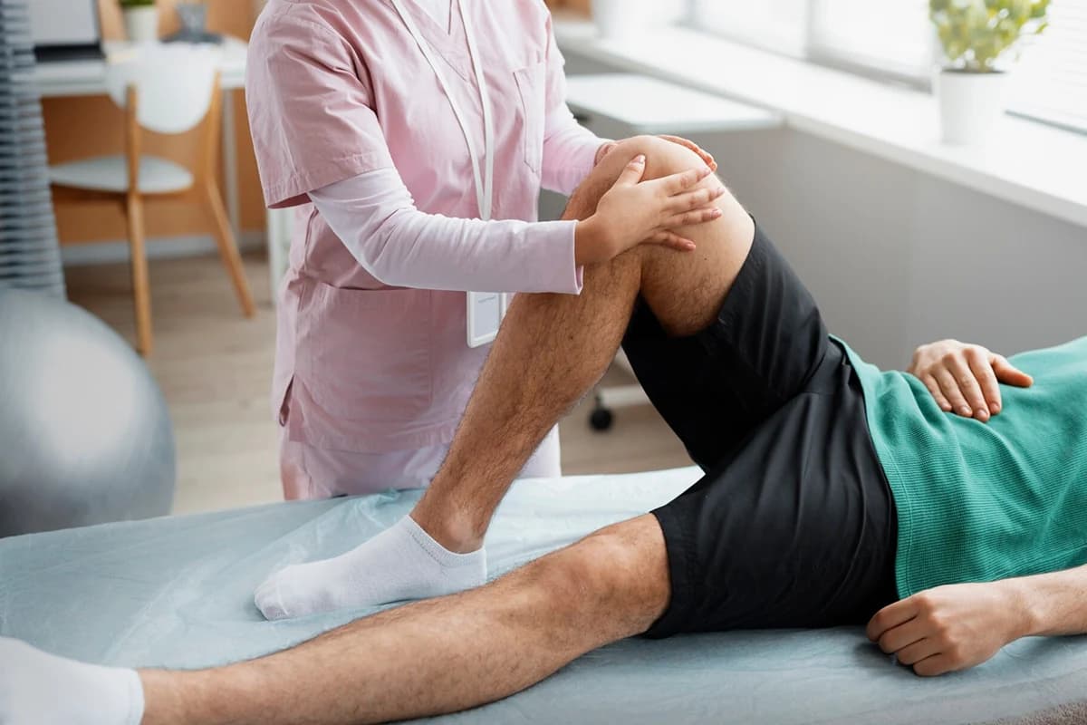 Knee Osteoarthritis (Gonarthrosis)
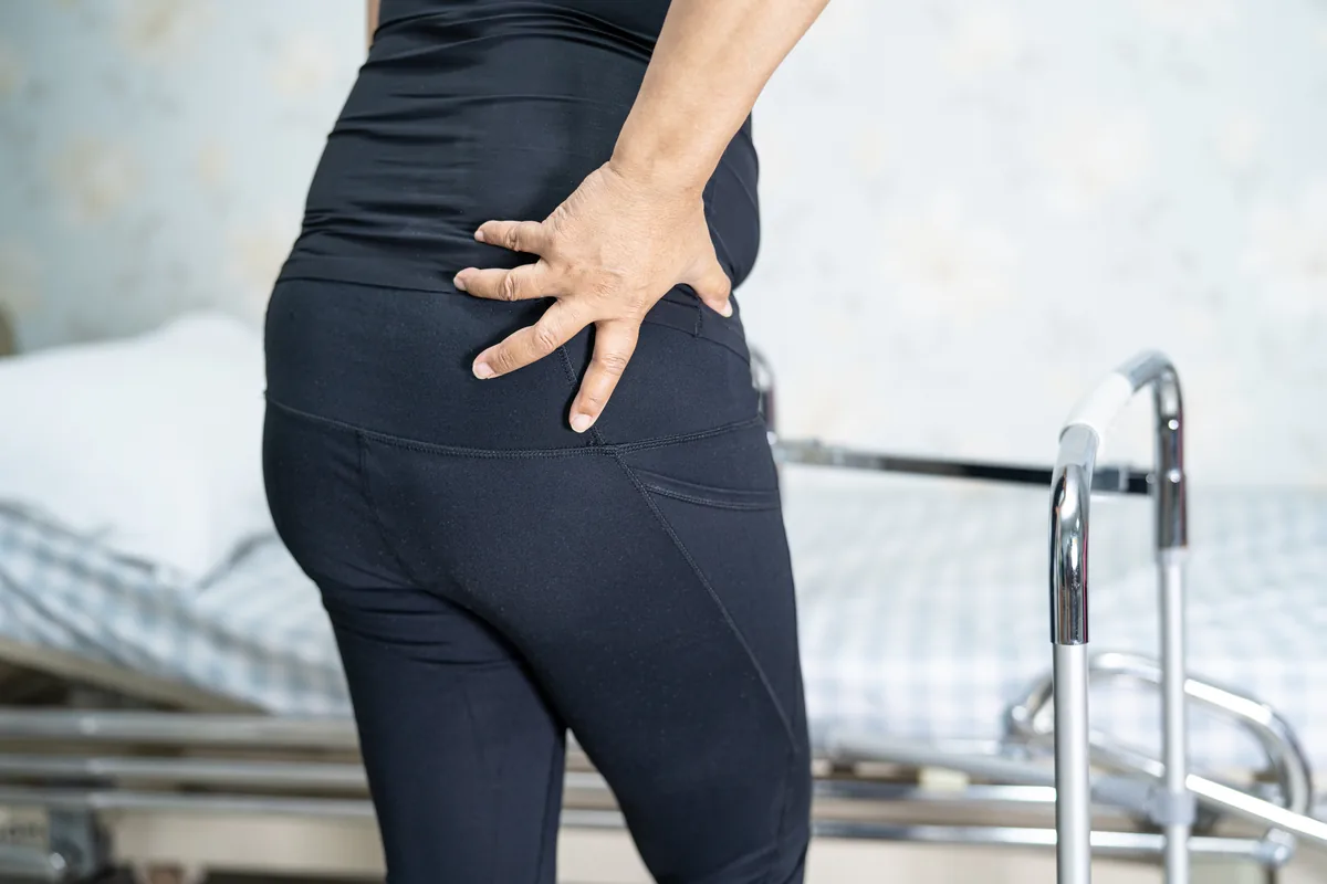 Hip Osteoarthritis (Coxarthrosis)
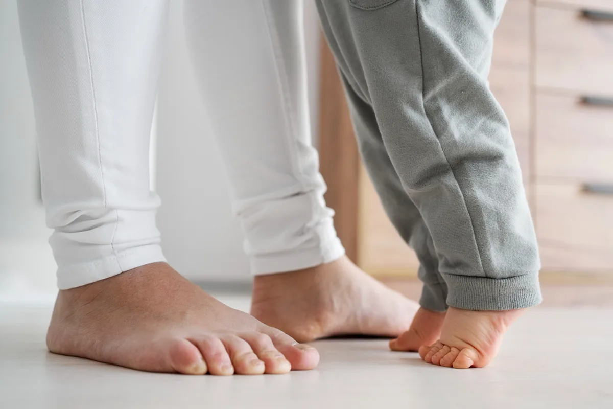 Toe Walking in Children
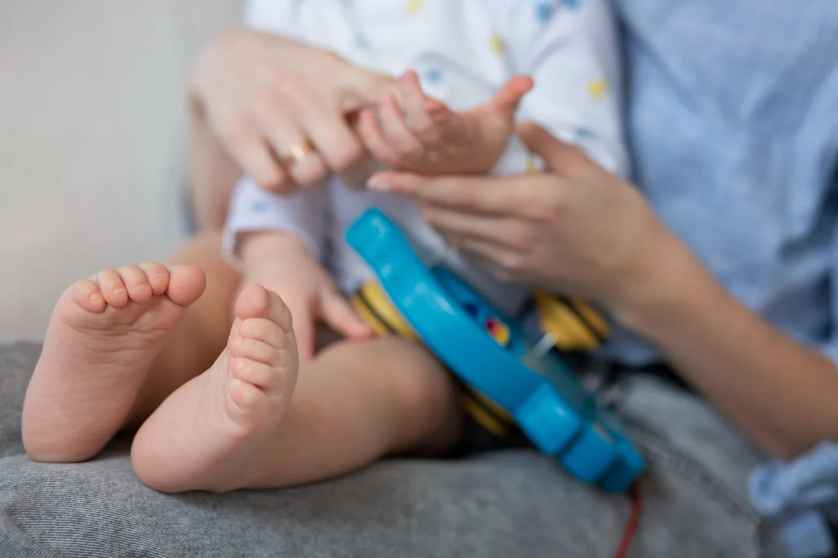 Clubfoot (Talipes Equinovarus)
Devices are used inside a programme and chosen after examination — they are not booked separately.
Your path to this treatment
Nothing here is booked from a page. You send the records you have, a rehabilitation physician reads them, and you get a written answer — including when the answer is that an intensive programme will not help.
4 steps, start to finish
1.Send your records
A discharge summary from the hospital that treated the original event, and any imaging of the affected area — CT, MRI or X-ray.2.A written plan in 48 hours
A rehabilitation physician reviews the case and returns the expected duration, the recommended programme and the centre. Free, no obligation.3.Travel and admission
Where a visa is needed we issue an invitation letter and treatment confirmation naming the dates and the facility. You are met at the airport and driven to the centre by private vehicle or ambulance; a bed for one companion and interpreting during therapy are arranged.4.Treatment, then home
The programme runs, then you leave with a discharge report and a written home exercise plan. Your return journey is organised before you go, with scheduled video reviews so progress does not stop.
In their words
What patients say after a programme
Short videos filmed during therapy: a child’s rehabilitation after a brain tumour, suspended gait training after spinal cord injury, and learning to walk again after a fall. Individual results vary.
Dave Evans on his physical therapy here
Musculoskeletal physiotherapy
Paediatric rehabilitation after a brain tumour
Inside a physiotherapy session
Spinal cord injury — the rehabilitation process
Suspension-assisted gait training
Walking difficulty and restricted movement after a fall
Regaining gait and range of motion
Locations
Centres that have this device — pick one.



Questions people actually ask
About this device
Who benefits from this?
Mainly children with cerebral palsy before surgical or botulinum decisions, and adults with complex gait problems after neurological injury or amputation.
How long does it take?
Around an hour including marker placement, longer in children who need time to become comfortable.
Is it uncomfortable?
No. Reflective markers are attached to the skin and you walk normally; nothing is invasive.
Do I receive the report?
Yes, in a form another clinician or surgeon can act on. That is the point of doing it.
Can a repeat analysis show whether treatment has worked?
Yes, provided it is repeated in the same way — the same laboratory procedure, footwear and walking aids — so that the two sessions are comparable. Only a change larger than the measurement error should be read as real: for most joint angles that error is under 5 degrees, and it is larger for hip and knee rotation 3.